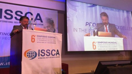
Bogota Course Will Share the Latest in Stem Cell Treatment Protocols
The two-day certification program is sponsored by ISSCA, will feature three topics new to the group’s Colombian training circuit
MIAMI LAKES, Florida— The International Society for Stem Cell Application (ISSCA), a global leader in stem cells research, applications, and education, has announced their latest certification program to be held in Bogota, Colombia on March 13-14, 2020.
The Stem Cells Certification course will feature a number of educational and hands-on sessions designed to give physicians the tools needed to integrate regenerative treatments into their practices. Registration for the two-day course is limited to eight participants to allow for intensive training and interaction.
During the two-day training, ISSCA faculty will share the latest stem cell protocols, including how to harvest and process stem cells derived from Stromal Vascular Faction (SVF) tissue and bone marrow.
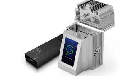
ISSCA faculty will additionally introduce three new protocols never before covered in past courses held in Colombia. The new curriculum includes clinical applications of allogeneic stem cells; using GCell technology, a device that can process SVF without enzymatic compounds; and an introduction to NK cell protocols, the newest regenerative medicine treatment to fight cancer.
“Our two-day training in Bogota will provide physicians with an invaluable opportunity to receive intensive hands-on training and education in some of today’s most effective and cutting-edge stem cells protocols,” said Benito Novas, ISSCA VP of Public Relations. “Physicians will learn from global leaders in the stem cells industry and be able to take practical application strategies back with them to integrate into their own practices to help patients suffering from degenerative diseases.”To learn more about the ISSCA, visit www.issca.us. More information about the Bogota certification course can be found at http://cursocelulasmadre.com/entrenamiento-bogota-programa/
- Published in Press Releases
Clinical trials on NK cells give hope for many people Who are suffering from cancer.
Induced pluripotent stem cells (also known as iPS cells or iPSCs) are a type of pluripotent stem cell that can be generated directly from a somatic cell. Pluripotent stem cells hold promise in the field of regenerative medicine. Because they can propagate indefinitely, as well as give rise to every other cell type in the body (such as neurons, heart, pancreatic, and liver cells), they represent a single source of cells that could be used to replace those lost to damage or disease.
Natural killer cells are the type of cytotoxic lymphocyte critical to the innate immune system. The role NK cells play is analogous to that of cytotoxic T cells in the vertebrate adaptive immune response. NK cells provide rapid responses to virus-infected cells, acting at around three days after infection, and respond to tumor formation.
Typically, immune cells detect the major histocompatibility complex (MHC) presented on infected cell surfaces, triggering cytokine release, causing apoptosis. NK cells are unique, however, as they can recognize stressed cells in the absence of antibodies and MHC, allowing for a much faster immune reaction.
Clinical Trial on NK cells
In a first clinical trial, a natural killer cell immunotherapy derived from induced pluripotent stem cells is being tested for safety in 64 patients with a variety of solid tumors. The first subjects used for the study received the cells in February at the University of California, San Diego (UCSD) Moores Cancer Center and MD Anderson Cancer Center.
This study is targeting late-stage cancer patients with solid tumors, including lymphoma, colorectal cancer, and breast cancer. The FT500 NK cells do not undergo any further alterations and after their derivation from the induced pluripotent stem cells (iPSCs), offering the possibility of a quicker, ready-made treatment.
Human embryonic stem cells induced iPSCs
Human embryonic stem cells (hESCs) and induced pluripotent stem cells (iPSCs) provide an accessible, genetically tractable, and homogenous starting cell population to efficiently study human blood cell development. These cell populations provide platforms to develop new cell-based therapies to treat both malignant and nonmalignant hematological diseases.
The NK cells are immune cells in the same family as T and B cells and are very good at targeting cancer cells for destruction. Some Laboratory experiments have shown they do so by attacking cells that have lost their significant self-recognition signals that tell the immune system not to attack. This is the phenomenon that can happen among cancer cells but not to healthy cells. Experts are not sure how many cancer cells lose that signal. Researchers are hopeful that the clinical trial can help determine which cancer patients could benefit the most from NK cell treatment.
iPS Clone
The ability to induce pluripotent stem cells from committed, human somatic cells provides tremendous potential for regenerative medicine. However, there is a defined neoplastic potential inherent to such reprogramming that must be understood and may offer a model for critical understanding events in the formation of the tumor. Using genome-wide assays, we identify cancer-related epigenetic abnormalities that arise early during reprogramming and persist in induced pluripotent stem cell (iPS) clones. These include hundreds of abnormal gene silencing events, patterns of aberrant responses to epigenetic-modifying drugs resembling those for cancer cells, and presence in iPS and partially reprogrammed cells of cancer-specific gene promoter DNA methylation alterations.
Progress in adoptive T-cell therapy for cancer and infectious diseases is hampered by the lack of readily available antigen-specific, human T lymphocytes. Pluripotent stem cells could provide an estimable source of T lymphocytes, but the therapeutic potential of human pluripotent stem cell-derived lymphoid cells generated to date remains uncertain.
Modification of T cells
Recently, some Approved cell therapies for Cancer also rely on modifying T cells, in those cases to produce cancer cell–binding chimeric antigen receptors (CARs), and have been effective in treating certain cancers such as leukemia.
Application of CAR T-Cell Therapy in Solid tumours
The Car T technology has wowed the field by all but obliterating some patients’ blood cancers, but solid malignancies present new challenges.
Therapies that contains such chimeric antigen receptor (CAR) T cells have been approved for some types of so-called liquid cancers of the blood and bone marrow, large B-cell lymphoma and B-cell acute lymphoblastic leukemia. But the approach has not had as much success for solid tumors.
Serious research into the therapy for brain cancer started almost 20 years ago after cancer biologist WaldemarDebinski, then at Penn State, discovered that the receptor for the immune signaling molecule interleukin 13 (IL-13) was present on glioblastomas, but not on healthy brain tissue. The receptor thus seemed like an excellent target to home in on cancer cells while sparing healthy ones. The CAR spacer domain that spans the immune cells’ membranes and its intracellular co-stimulatory areas, as well as the process used to expand cells outside the body, to boost the T cells’ activity.
CAR T- A Safer Cell Therapy
While managing CAR T-cell therapy toxicity could help keep already-designed treatments on their march to the clinic, many immunotherapy companies are also working to develop a new generation of inherently safer therapies, yet just as efficient. A crucial part of achieving this goal will be improving CAR specificity for target cells. With current treatments, the destruction of normal cells is often an unavoidable side effect when healthy tissue carries the same antigens as tumors; noncancerous B cells, for example, are usually casualties in CD19-targeted therapies.
CAR T delivery is a non-easy factor in the treatment of solid tumors and other unknown forms of tumors. With the non-solid cancers, cells are administered by a blood infusion, and once in circulation, the CAR T can seek out and destroy the rogue cells. For solid tumors, it’s not so simple.
The main drawback of taking cells from a patient and developing them into a cellular immunotherapy product is that the process can take weeks.
Patel tells The Scientist “But for the majority of patients who may not be a candidate or may not have time to wait for such an approach, the idea that there’s off-the-shelf immunotherapy that could potentially as a living drug act against their cancer, I think is a fascinating concept,”
- Published in Corporate News / Blog
Stem cell treatment could offer one-end-solution to Diabetes
Insulin-producing cells grown in the lab could provide a possible cure for the age-long disease (diabetes).
Type 1 diabetes is an auto¬immune disease that wipes out insulin-producing pancreatic beta cells from the body and raises blood glucose to dangerously high levels. These high levels of Blood sugar level can be even fatal. Patients are being administered insulin and given other medications to maintain blood sugar level. To those who cannot maintain their blood sugar level, they are given beta-cell transplants but to tolerate beta cell transplants; patients have to take immunosuppressive drugs as well.
A report by a research group at Harvard University tells us that they used insulin-producing cells derived from human embryonic stem cells (ESCs) and induced pluripotent stem cells to lower blood glucose levels in mice. Nowadays, many laboratories are getting rapid progress in human stem cell technology to develop those cells that are functionally equivalent to beta-cells and the other pancreatic cell types. Other groups are developing novel biomaterials to encapsulate such cells and protect them against the immune system without the need for immunosuppressant.
Major pharmaceutical companies and life sciences venture capital firms have invested more than $100 million in each of the three most prominent biotechnological industries to bring such treatments into clinical use:
- Cambridge
- Massachusetts–based companies Semma Therapeutics
- Sigilon Therapeutics, and ViaCyte of San Diego
Researchers of UC San Francisco have transformed human stem cells into mature insulin-producing cells for the first time, a breakthrough in the effort to develop a cure for type-1 (T1) Diabetes. Replacing these cells, which are lost in patients with T1 diabetes, has long been a dream of regenerative medicine, but until now scientists had not been able to find out how to produce cells in a lab dish that work as they do in healthy adults.
What is T1 diabetes?
T1 diabetes is an autoimmune disorder that destroys the insulin-producing beta cells of the pancreas, typically in childhood. Without insulin’s ability to regulate glucose levels in the blood, spikes in blood sugar can cause severe organ damage and eventually death. The condition can be managed by taking regular shots of insulin with meals. However, people with type 1 diabetes still often experience serious health consequences like kidney failure, heart disease and stroke. Patients facing life-threatening complications of their condition may be eligible for a pancreas transplant from a deceased donor, but these are rare, and they are supposed to wait a long time.
Researchers have just made a breakthrough that might one day make these technologies obsolete, by transforming human stem cells into functional insulin-producing cells (also known as beta cells) – at least in mice.
“We can now generate insulin-producing cells that look and act a lot like the pancreatic beta cells you and I have in our bodies,” explains one of the team, Matthias Hebrok from the University of California San Francisco (UCSF).
“This is a critical step towards our goal of creating cells that could be transplanted into patients with diabetes.”
Type-1 diabetes is characterized by a loss of insulin due to the immune system destroying cells in the pancreas – hence, type 1 diabetics need to introduce their insulin manually. Although this is a pretty good system, it’s not perfect.
Making insulin-producing cells from stem cells
Diabetes can be cured through an entire pancreas transplant or the transplantation of donor cells that produce insulin, but both of these options are limited because they rely on deceased donors. Scientists had already succeeded in turning stem cells into beta cells, but those cells remained stuck at an early stage in their maturity. That meant they weren’t responsive to blood glucose and weren’t able to secrete insulin in the right way.
Scientists at the University of California San Francisco made a breakthrough in the effort to cure diabetes mellitus type 1.
For the first time, researchers transformed human stem cells into mature insulin-producing cells, which could replace those lost in patients with the autoimmune. There is currently no known way to prevent type-1 (T1) diabetes, which destroys insulin production in the pancreas, limits glucose regulation, and results in high blood sugar levels. The condition can be managed with regular shots of insulin, but people with the disease often experience serious health complications like kidney failure, heart disease, and stroke.
“We can now generate insulin-producing cells that look and act a lot like the pancreatic beta cells you and I have in our bodies,” according to Matthias Hebrok, senior author of a study published last week in the journal Nature Cell Biology.
“This is a critical step toward our goal of creating cells that could be transplanted into patients with diabetes,” Hebrok, director of the UCSF Diabetes Center, said in a statement.
Islets of Langerhans are groupings of cells that contain healthy beta cells, among others. As beta cells develop, they have to separate physically from the pancreas to form these islets.
The team artificially separated the pancreatic stem cells and regrouped them into these islet clusters. When they did this, the cells matured rapidly and become responsive to blood sugar. In fact, the islet clusters developed in ways “never before seen” in a lab. After producing these mature cells, the team transplanted them into mice. Within days, the cells were producing insulin similar to the islets in the mice. While the study has been successful in mice, it still needs to go through more rigorous testing to see if it would work for humans as well. But the research is up-and-coming. “We can now generate insulin-producing cells that look and act a lot like the pancreatic beta cells you and I have in our bodies. This is a critical step towards our goal of creating cells that could be transplanted into patients with diabetes,” He said.
“We’re finally able to move forward on several different fronts that were previously closed to us,” he added. “The possibilities seem endless.”
Basic research keeps elucidating new aspects of beta cells; there seem to be several subtypes, so the gold standard for duplicating the cells is not entirely clear. Today, however, there is “a handful of groups in the world that can generate a cell that looks like a beta cell,” says Hebrok, who currently acts as scientific advisor to Semma and Sigilon, and has previously advised ViaCyte. “Certainly, companies have convinced themselves that what they have achieved is good enough to go into patients.”
The stem cell reprogramming methods that the three companies use to prompt cell differentiation create a mixture of islet cells. Beta cells sit in pancreatic islets of Langerhans alongside other types of endocrine cells. Alpha cells, for example, churn out glucagon, a hormone that stimulates the conversion of glycogen into glucose in the liver and raises blood sugar. Although the companies agree on the positive potential of islet cell mixtures, they take different approaches to developing and differentiating their cells. Semma, which was launched in 2014 to commercialize the Harvard group’s work and counts Novartis among its backers, describes its cells as fully mature, meaning that they are wholly differentiated into beta or other cells before transplantation. “Our cells are virtually indistinguishable from the ones you would isolate from donors,” says Semma chief executive officer BastianoSanna
To get around the donor problem, researchers, including the team at UCSF has been working on nudging stem cells into becoming fully-functional pancreatic beta cells for the last few years. Still, there have been some issues in getting them all the way there.
“The cells we and others were producing were getting stuck at an immature stage where they weren’t able to respond adequately to blood glucose and secrete insulin properly,” Hebrok said.
“It has been a major bottleneck for the field.”
“We’re finally able to move forward on a number of different fronts that were previously closed to us,” Hebrok added. “The possibilities seem endless.”
Regardless of starting cell type, the companies say they are ready to churn out their cells in large numbers. Semma, for example, can make more islet cells in a month than can be isolated from donors in a year in the United States, Sanna says, and the company’s “pristine” cells should perform better than donor islets, which are battered by the aggressive techniques required for their isolation.
As these products, some of which have already entered clinical trials, move toward commercialization, regulatory agencies such as the US Food and Drug Administration (FDA) and the European Medicines Agency have expressed concern about the plasticity of the reprogrammed cells. All three firms subject their cells to rigorous safety testing to ensure that they don’t turn tumorigenic. Before successful trials, companies won’t know the dose of beta cells required for a functional cure, or how long such “cures” will last before needing to be boosted. There’ll be commercial challenges, too: while the companies are investing heavily to develop suitable industrial processes, all acknowledge that no organization has yet manufactured cell therapies in commercial volumes.
Nevertheless, there’s growing confidence throughout the field that these problems will be solved, and soon. “We have the islet cells now,” says Alice Tomei, a biomedical engineer at the University of Miami who directs DRI’s Islet Immuno-engineering Laboratory.
“These stem cell companies are working hard to try to get FDA clearance on the cells.”
Protecting stem cell therapies from the immune system
Whatever the type of cell being used, another major challenge is delivering cells to the patient in a package that guards against immune attack while keeping cells fully functional. Companies are pursuing two main strategies:
- Microencapsulation, where cells are immobilized individually or as small clusters, in tiny blobs of a biocompatible gel.
- Macroencapsulation, in which greater numbers of cells are put into a much larger, implantable device.
ViaCyte, which recently partnered with Johnson & Johnson, launched its first clinical trial in 2014. The trial involved a micro-encapsulation approach that packaged up the company’s partially differentiated, ESC-derived cells into a flat device called the PEC-Encapsulation. About the size of a Band-Aid, the device is implanted under the skin, where the body forms blood vessels around it. “It has a semipermeable membrane that allows the free flow of oxygen, nutrients, and glucose,” says ViaCyte’s chief executive officer, Paul Laikind. “And even proteins like insulin and glucagon can move back and forth across that membrane, but cells cannot.”
The trial showed that the device was safe, well-tolerated, and protected from the adaptive immune system—and that some cells differentiated into working islet cells. But most cells didn’t engraft effectively because a “foreign body response,” a variant of wound healing, clogged the PEC-Encap’s membrane and prevented vascularization. ViaCyte stopped the trial and partnered with W. L. Gore & Associates, the maker of Gore-Tex, to engineer a new membrane. “With this new membrane,” says Laikind, “we’re not eliminating that foreign body response, but we’re overcoming it in such a way that allows vascularization to take place.” The company expects to resume the trial in the second half of this year, provided it receives the green light from the FDA.
Semma is also developing macro¬-encapsulation methods, including a very thin device that in prototype form is about the size of a silver dollar coin. The device is “deceptively simple, but it allows us to put [in] a fully curative dose of islets,” Sanna says.
Semma is also investigating microencapsulation alternatives. At the same time, the company is advancing toward clinical trials using established transplantation techniques to administer donated cadaver cells to high-risk patients who find it particularly difficult to control their blood glucose levels. These cells are infused via the portal vein into the liver, and patients take immunosuppressive drugs to prevent rejection.
Sigilon is working on its microencapsulation technology. Launched in 2016 on the back of work by the labs of Robert Langer and Daniel Anderson at MIT, the company has created 1.5-millimeter gel-based spheres that can hold between 5,000 and 30,000 cells (Nat Med, 22:306–11, 2016). Each sphere is like a balloon, with the outside chemically modified to provide immune-protection, says Sigilon chief executive officer Rogerio Vivaldi. “The inside of the balloon is full of a gel that creates almost a kind of a matrix net where the cells reside.”
In 2018, shortly after partnering with Eli Lilly, Sigilon and collaborators published research showing that islet cells that were encapsulated in gel spheres and transplanted into macaques remained functional for four months. The company has not disclosed a time frame for a type 1 diabetes trial “but we’re moving pretty quickly,” says chief scientific officer David Moller.
Conclusion
To conclude, all three firms hope to extend their work to treat some of the 400 million people worldwide with type 2 diabetes, many of them eventually benefit from insulin injections. The recent endorsements from big Pharmaceutical underline the real progress in beta-cell transplants, says Aaron Kowalski, a molecular geneticist and chief executive officer at JDRF, a foundation based in New York that has funded research at ViaCyte and academic labs whose work has been tapped by Semma and Sigilon. “These companies all realize that if they don’t do it, somebody else will. It’s hard to predict exactly when, but somebody is going to make this work.”
- Published in Corporate News / Blog
Bats Carry Corona Virus. So Why Don’t They Get Sick?
A lot of viruses that has taken a toll on life, the ebola virus in Africa, The Nipah virus of Nipah and the most recent one corona virus that left china running helter skelter all seemed to have originated from bats. During the course of the virus epidemic in Wuhan where it was first detected, some Chinese researchers in Wuhan examined some patients affected in that area and then took samples of the virus.
They did findings on the genetic sequence of the virus with other viruses that were known. The corona virus surprisingly had a 96% match with the horseshoe bats that are dominant in the southwest of china. The research findings were then published in a study on February, 2020.
A virologist Vineet Menachery from the university of Texas Medical Branch at Galveston though not affiliated to the study said “They’re too close in terms of their pure genetics to say they’re not related, or that they didn’t have a common ancestor.”
Menachery was a reputable virologist and had done other research works. He contributed to the theory that the spread of the corona virus must have been from these bats to humans. And possibly must have had another animal that served as an intermediary for the spread.
This same thing had happened with other forms of corona viruses as noted in the case of SARS (Severe Acute Respiratory Syndrome) an outbreak that took place in 2002-2003 where civets, a mongoose family member were infected with the bat corona virus and spread as humans bought them for food.
Another case was the MERS (Middle East Respiratory Syndrome) outbreak. This one happened in 2012 and was as a result of infected camels from the virus. People who ate undercooked meat of camels and as well drank the raw milk of camels were all affected.
So why is it that there are so many diseases that are spread from bats?
Its no doubt, bats have a lot of viruses that they carry with them. And these viruses in their variety are spread and manifests its tolls on people. Scientist are not sure why this is the case as confirmed by Kevin Olival, a research vice president as EcoHealth Alliance, a non-profit organization based in the U.S. He went further to say that it may have something to do with the family of the viruses carried by the bats. So you know, there are over 130 different families of viruses that bats do carry around.
And then, most bats and humans do come in contact through several means. The millions of populations of bats are ubiquitous to all the continents apart from in antartica. Rebekah of Colorado State university who researched infectious pathogens said “There’s a lot of viruses we’re finding in bats because there’s a lot of bats out there.”
They move about in multitudes and live in colonies of large populations. Some these members live in caves and share caves and trees where there can be a contact between humans and bats. Hence, these viruses can spread from these bats to humans.
Despite their sizes, bats have relatively long lifespans and can live over 30 years.”So there’s a long time for them to be persistently infected with the virus and shed it into the environment,” Kading says. The mode of mechanisms for these viruses are through urine, saliva and feces of bats. The outbreak of Nivah that happened in Bangladesh was linked to the sap of a date palm gotten from some trees that some bats licked and had infested with their urine.
Reading through all these, it is not absurd to wonder why the bats themselves do not get affected by the viruses they carry.
The answer to that question is based on the fact that the bat is the only flying mammal in the world. Their body metabolism and process quite differ from that of normal mammals too. When bats fly, their heart rates rise to about a thousand beats per minute with a temperature rise of about 100 degrees Fahrenheit. Linfa Wang a student of bat viruses at Duke-NUS Medical School in Singapore says that when these signs manifest in other mammals, they are signals that can trigger death. But this is not the same case for other bats. This is a lifestyle for them, every day.
Their system is also capable of producing molecules that other organisms do not have. The molecules carry out repair functions and prevent cell damage. This makes their system a bit irresistible to infections and also make them recalcitrant to viruses and resilient to diseases such as diabetes, cancer and other health conditions.
This is a prove that the manifestation of viruses in mammals is not always as a result of the virus itself, but as a result of the body’s reaction to the presence of such a virus that makes us ill by triggering other chain reactions, as Wang explains.
Olival at EcoHealth explains that these bats have coevolved with these viruses and it is not totally their fault that we humans are infected and affected by these viruses. The actual problem is when the viruses move from their species to other species of mammals which is also fostered by human activity.
Naturally, it would be hard for most animals and mammals to cross paths. But Olivial says that the presence of some activities and availability of exchange platforms made available by humans can allow such interaction to occur. She gave an example using wildlife markets like the one in Wuhan, where a bat could be mixed up with a civet. Who later on come in contact with humans – eg. Butchers who do not observe proper hygiene and protection from animal blood.
“The way that we’re coming into contact with these animals, hunting, selling, and trading them is to a scale that really we haven’t seen before,” he says.
Investigative teams did some in-depth search and they discovered some traces of this virus in 22 stalls and in a garbage truck that was found at Huanan Seafood Market right there in Wuhan, a place known for booming trade for live animals. This discovery led to shutting down the market as it was tied to majority of the cases.
The intermediary animals to this viruses are still a mystery, but it is clear that some of these animals are prone to interact more with humans. This is why when they are infected, the likelihood for human infection is widened. These other infected animals can sneeze, urinate, be cooked as food or even owned as pets.
Bats are not just vectors for viruses, they play an important role in balancing the eco-system. They feed on insects and fruits and are active agents of pollination. In fact, Wang believes that since these bats have successfully coevolved with these viruses, there is every possibility that they can be the agents that can lead to the cure and provision of therapies for these viruses.
- Published in Corporate News / Blog
Mexico City Medical Congress to Showcase the Global Stem Cells Group’s Latest Innovations
The group is sponsoring the event, which will attract physicians from across the globe with an interest in longevity and anti-aging medicine
MIAMI LAKES, Florida— The Global Stem Cells Group (GSCG) is set to sponsor the XI Congreso Mundial de Medicina Antienvejecimiento y Longevidad (World Conference of Anti-Aging and Longevity Medicine) to be held in Mexico City, Mexico on February 16-18, 2020.
The medical congress is expected to attract over 450 physicians and researchers from across the world interested in anti-aging and longevity practices and medical innovations. Over 30 speakers are slated to share information with attendees on a wide range of topics on how to lead a long, healthy life and improve longevity.
The GSCG is set to share a number of its latest innovations with congress attendees, including its newly released GCell technology device. This cutting-edge tool utilizes micrograft technology to harness the natural and powerful restorative capabilities of adipose tissues. Because it is FDA compliant, the device allows physicians across the globe to continue practicing adult stem cells-based procedures.
Additional benefits of GCell technology include shorter treatment times, delivering in-office treatments in around 30 minutes with local anesthesia, as well as less fat collection compared to existing treatments (15 mL versus 50 mL). GCell technology holds exciting implications across a range of medical specialties, including orthopedics, dermatology, cosmetic gynecology, aesthetics, and hair loss.
In addition to its GCell technology, the GSCG will also feature its newest line of stem cells products derived from first-tissue exosomes. Cellgenic Flow Exosomes utilizes the latest science and research available in cellular therapies to deliver a non-surgical approach to creating regenerative responses in a broad range of treatments. The product utilizes exosomes, which replicate the signals given out by stem cells, versus actual stem cells. Exosomes play a pivotal role in cell-to-cell communication and are involved in a wide range of physiological processes. These particles transfer critical bioactive molecules such as proteins, mRNA, and miRNA between cells and regulate gene expression in recipient cells.
“The XI Congreso Mundial de Medicina Antienvejecimiento y Longevidad is one of the world’s premier events connecting physicians and researchers with today’s most innovative treatments and technologies utilizing regenerative medicine,” said Benito Novas, CEO of the GSCG. “As a worldwide leader in training, education, and innovative products in the field of regenerative medicine, the GSCG is pleased to sponsor this congress and share its exciting new portfolio of products with physicians from across the world.”
To learn more about the Global Stem Cells Group and all of the group’s latest news and innovations, visit http://www.stemcellsgroup.com/
- Published in Press Releases
Two Global Stem Cells Group Officials to Speak at Paraguay Conference
The group is set to sponsor the 2nd Congress of Aesthetic Medicine and Regenerative Gynecology to be held in Paraguay
MIAMI LAKES, Florida—The Global Stem Cells Group (GSCG) has announced its sponsorship of the 2nd Congress of Aesthetic Medicine and Regenerative Gynecology to be held on March 12-13, 2020 in Asuncion, Paraguay. The GSCG’s CEO Benito Novas and Development and Research Director Dra. Maritza Novas will each deliver key lectures during the annual medical congress, which will focus on advancements in the fields of aesthetic medicine and regenerative gynecology.
Benito Novas serves as the GSCG’s CEO and will lead a lecture on regenerative medicine marketing strategies. His talk will give practical marketing tools and advice to physicians running their own private practices in aesthetic medicine. In addition to his role as CEO with the GSCG, Novas also serves as the Head of Public Relations for the International Society for Stem Cell Application (ISSCA) and has authored two books on marketing strategies for clinics in the regenerative medicine industry, Your Aesthetic Practice: What Your Patients Are Saying and Marketing Digital en Su Clínica Estética (https://www.thriftbooks.com/a/benito-novas/2752773/).
Dra. Maritza Novas serves as the GSCG’s Development and Research Director as well as the US Director for the ISSCA. She will deliver the opening lecture for the congress, discussing clinical applications of regenerative medicine in the aesthetic field. Her lecture will be informed by clinical case studies demonstrating the efficacy of regenerative treatments in aesthetics. Dra. Novas is well-renowned in the field of regenerative medicine for making a profound impact on her patients through innovative treatment protocols. She has additionally trained countless doctors looking to add regenerative medicine to their practices.
On March 14, following the conclusion of the congress, representatives from the GSCG will also offer a certification course in cellular therapy. The course is designed to help all physicians who want to add stem cells therapies based on birth tissue derived components to their practices. Attendees will learn about the latest stem cell products, such as exosomes, cord blood, and amniotic fluid. Additional topics to be covered will include how to choose the right products to treat specific conditions and details about how to handle the manufacturing process, from selecting a healthy donor through quality control and storage at the medical office.
“The Global Stem Cells Group is pleased to serve as a sponsor for the 2nd Congress of Aesthetic Medicine and Regenerative Gynecology,” said Benito Novas. “We look forward to sharing today’s best research on marketing strategies to help regenerative medicine practices grow their success and influence as well as sharing clinical applications of stem cells treatments in aesthetic medicine. As a global leader in stem cells education, we always look forward to these events to help more physicians gain the knowledge and skills necessary to bring life-saving treatments to more patients across the globe.”
To learn more about the Global Stem Cells Group and all of the group’s latest news and innovations, visit http://www.stemcellsgroup.com/.
- Published in Press Releases
Global Stem Cells Group to Sponsor Miami Congress on Anti-Aging and Longevity
The group will launch its innovative GCell technology and a new online course at the event, while CEO Benito Novas will lead a lecture about marketing in the regenerative medicine field
MIAMI LAKES, Florida—The Global Stem Cells Group (GSCG) is set to sponsor the Second Intercontinental Medical Congress on Anti-Aging and Longevity (https://miami2020.semal.org/) hosted by the Spanish Society of Anti-Aging Medicine (SEMAL) and the Federation of Latin American of Anti-Aging Medicine (FISMAL). The congress will be held on February 6-9, 2020 in Miami, Florida at the Hampton Inn & Suites by Hilton Miami Brickell.
During the event, the Global Medical Group, a subsidiary of GSCG, will launch its GCell micrograft technology, an autologous tissue suspension that boasts minimal manipulation. The product has application implications for a wide range of medical uses, including orthopedics, dermatology, cosmetic gynecology, aesthetics, and hair loss.
GCell technology will allow physicians utilizing regenerative medicine to continue practicing adult stem cells-based procedures thanks to the product’s adherence to FDA compliance standards since the procedure does not utilize enzymes.
GCell technology is a closed-system medical device that harnesses the natural and powerful restorative capabilities of adipose tissue. The GCell SVF (Stromal Vascular Fraction) procedure can be performed in office in around 30 minutes, while existing procedures can take up to two hours. It also utilizes only local anesthesia, and performing physicians only have to collect 15 ml of fat instead of the 50 ml required for traditional protocols.
During the congress, the GSCG will also be launching its new online stem cells course, which will benefit doctors interested in learning about the newest stem cells protocols and technologies. The course will specifically focus on stem cells protocols based on first tissue derived compounds, such as exosomes, cord blood, and amniotic fluid.
In addition to launching GCell technology and its new course, the GSCG’s CEO and founder Benito Novas will be giving a lecture in the main room. He will be speaking to attendees about the newest challenges facing the regenerative medicine filed and what types of marketing strategies practitioners should implement to run a successful practice integrating regenerative medicine.
“The Global Stem Cells Group is looking forward to attending the Second Intercontinental Medical Congress on Anti-Aging and Longevity to talk with attending physicians and researchers about the technological and educational advances we are making to help move the field of regenerative medicine forward,” said Novas.
To learn more about the Global Stem Cells Group and all of the group’s latest news and innovations, visit http://www.stemcellsgroup.com/.
- Published in Press Releases
A New Medical Device, in the Management of Complex Wounds
Because of the complex nature of wound healing process, an injury on the skin can pose several challenges and are likely pose complications especially when they are acute. They can as well deteriorate from acute to chronic conditions which will require external intervention best understood by a specialist physician to get the area affected by the wound under normalcy.
The complexity of wound healing and research remains an ocean of knowledge that is continuously researched intensely to uncover depths of wound healing techniques and interventions. Hence, this report contains an introduction and details to the use of a new medical innovation called Gcells used primarily for the management of wounds in their different etiology.
In a case where the process of wound healing seemed difficult, Gcells proved great effects an attribute to their design and working protocol. Gcells are conditioned to work with an enriched suspension of progenitor cells that can efficiently aid tissue repair process. In this case report, two subjects were used as donors and acceptors of these micro-grafts.
Introduction
The skin is an outer layer of the body, offering protection to the underlying layers. A wound breaks this layer and inhibits the various functions as well as expose or also break the underlying layer of tissues. Repair processes are inherent and part of homeostatic processes of the body to try to restore the skin back to its normalcy in structure and in function.
The basics for the skins repair mechanism is represented by a cloth and an inflammation where vessels dilate and monocytes activate leading to breakdown of necrotic tissues. This basic process can be inhibited or delayed by a number of varying factors that lead to deteriorative transformation of acute wounds to chronic forms. But if there is no alteration in the repair process, Mesenchymal cells kickstart proliferative process and begin to repair and restructure the affected tissues starting from the base. At the same time epithelial tissues begin to grow around the wound leading to a final step of the healing process. In this final stage, remodeling of the skin structure is primary and then maturation of a scar.
These processes are efficient best in certain conditions which if affected by factors such as cardiovascular ailment, diabetes, bacterial or any other genre of infection, can inhibit these processes.
Hence, it is necessary to understand in details these processes if there is going to be development or innovation for effective healing processes. Just as stated above, during the proliferative phase of wound healing, Mesenchymal cells are the key role players. Their structure includes a Mesenchymal stem cell (MSCs), multi potent in nature and offer supportive, therapeutic and trophic functions. They are also able to release viable trophic, anti-inflammatory cytokines and anti-apoptotic molecules that offer protection during the repair of wounded skin. MSCs also possess subpopulations that are stem-like nature commonly referred to as “side population” (SP) they have been found out to be enriched in over 1000-fold of progenitor cells and multipotent stem cells and as well exist in tissues and tumors. SP exists in a variety of organs and tissues, after an original discovery to be prominent in the bone marrow of a mouse. The organs with SP include the lung, liver, brain, mammary gland and in skeletal muscles.
In other discoveries, it was discovered that they probably may also be isolated in other tissues of the body. This discovery was in an in vitro and in vivo experiment when Dental Pulp Stem Cells (DPSCs) showed capability to differentiate into osteoblasts and built a woven bone by forming an Extracellular Matrix (ECM) secreted by the osteoblasts. The experiment drew results on the both the quality and quantity of the matrix formed by the DPSCs in the in vivo and in vitro experiment using Stem cells and accompanying biomaterials.
Thus proved that dental pulp holds potentialities of therapeutic strength and a rich source of progenitor/autologous cells that can be used to aid healing processes even applicable to regeneration of craniofacial bones.
This is the evidence that supports the working principle of Gcells innovation. Gcell successfully separates this side population with a size of 50 micron. At this cell population, they can form autologous micro-grafts and can either be used alone or alongside biomaterials prepared in a biocomplex ready for use when necessary.
In this case report, two subjects were used as donors and acceptors of these micro-grafts for enhanced healing of complex wounds through autologous micro-grafts using the Gcell.
Clinical case 1
The first case involves a woman at age 50 who does not have any diseases or disorders. She underwent a laparoscopic gastric bypass surgery and was doing well considering parameters of weightloss. Two years later she moved in for abdominoplasty bariatric. Later on, post complications showed preeminence of necrosis which was discovered after first medical examination were about 150 to 200cm2 at the end of the flaps. An initial necrosectomy showed an intense loss of tissue and we furthered to place the wound on VAC therapy and the patient in active participation of this therapy for one week then at home as an outpatient.
As an outpatient, there was improved and progressive wound cleansing while granulated tissues around the base area were cleared.
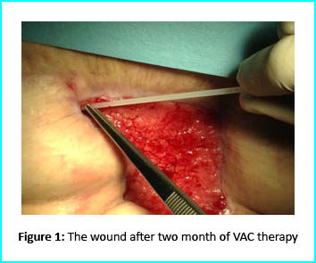
The VAC therapy after 2 months still left the margins of the wound deteriorated and surrounding areas not in axis with skin surface.
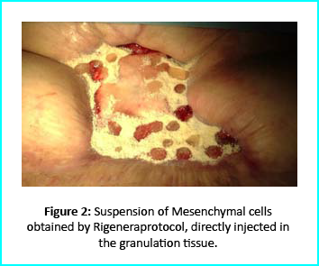
The Gcell protocol kickstarted after consent from the outpatient. We started by collecting a 3 cm2 skin sample from the patient for the purpose of obtaining the cell suspension needed to be injected to the granulation tissue (figure2).
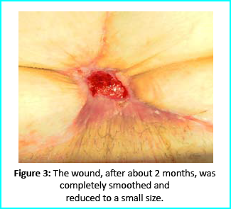
We followed up with conventional wound treatment as in cleaning and replacement with sterile gauze dabbed with Vaseline. The wound area began to improve in both healing progress and general appearance. In two months, the undermined area disappeared as well as leveled to the axis of the skin surface. 2 months later, the wound reduced to a very little scar that is mild and smoothed compared to the initial condition. (figure 3).
A man who suffered liver cirrhosis, hiatal hernia and diabetic as well at the age of 78. Complex surgery was carried out and distal esophagectomy was performed. But hiatal hernia was not decreased into the abdomen, so he was booked up for corrective surgery. During the intervention the adhesions correlated to the previous abdominal operation and led to opening the colon for resection. Some postoperative complications by the appearance of entero-cutaneous fistulas, related to a colonic anastomosis dehiscence. A second intervention was inevitable hence a ileostomy protection and repackaging of colonic anastomosis. We closed the laparotomy using a biological prosthesis. But we met further complications from ascetic failure that needed intensive care hepatology.
Patient’s condition that included poor liver synthesis had its toll on the healing of the surgical wound. Just as the first case, necrotic tissues grew around the biological prosthesis. We conducted necrosectomy and the biological prosthesis was left half exposed. (Figure 5).
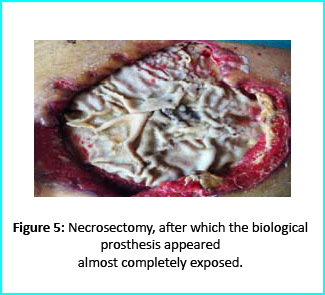
Further treatment of the wound using advanced medication helped cover the biological prosthesis with granulation tissue (figure 6).
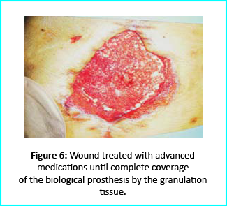
Plastic surgeons conducted evaluations on the patient and the choice to do a rotation flap did not seem so appropriate. VAC therapy was used on the wound for about 15 days even though the device wasn’t efficient enough to maintain supposed suction in the presence of ileostomy. We proceeded to treat the patient further with Gcell protocol when wound dimension progressed to about 250cm2. The tissue granulation was of right margin near the ileostomy improved even though it appeared to be undermined. In summary, Gcell protocol has proved a great level of efficiency in healing and restoration of damaged tissues. This progress is certain to open way for employment in the clinical practice that involves the treatment and management of acute and chronic wounds and in any other field of medicine that will inevitably need an instrument to repair lesion on tissues.
Discussion and conclusion
We made it clear earlier in this document about the efficiency of Gcell protocol in its aid to wound healing especially for wounds that are likely to develop from acute to chronic conditions. The working principle for the Gcell used to obtain the viable progenitor cells used for the micrografts relies on one individual as both the donor and the acceptor. This will help to reduce complications that are related to implants or injected micrografts that are non-autologous. Gcell is flexible and can be used both during in operating rooms as well as in ambulatories. This innovation is vastly spreading and currently used in the fields of oral-maxillo-facial field proven by recent studies even though a greater area of its application widespread and acceptable in plastic surgery, dermatology and orthopedics.
Conclusion of this report brings to clarity in demonstration, an efficient, useful and low-risk innovation in the field of medicine, useful for areas in wound management and healing. However, the viability of the Gcell product still needs to be texted on subjects with different conditions and perspectives. But we assure that this device will prove to be a better therapeutic approach in the field of medicine in improving healing of complex wounds. This confidence lies in the excellent features and working principles of this device in obtaining cell suspension, flexibility, facility for procedure and more importantly, the cost. This will help reduce the use of exorbitantly prices devices for advanced medication. In summary, apart from introducing an efficient innovation in the medicine. Gcell has the potentialities to offer employment on clinical procedures that will help aid in the management of wounds no matter how the case may be.
- Published in Corporate News / Blog
Global Stem Cells Group Enters Training Agreement with Moroccan Hospital
The agreement between Global and CHU Mohammed VI in Marrakech will help enhance staff preparation and clinical offerings in regenerative medicine
MIAMI LAKES, Florida— Stem Cells Training Inc., a subsidiary of the Global Stem Cells Group (GSCG), has announced that it has signed an onsite training agreement with Centre Hospitalo-Universitaire Mohammed VI Marrakech (CHU). GSCG faculty will travel to Marrakech, Morocco to provide this invaluable two-day training to CHU’s staff on February 14 and 15, 2020.
The agreement names the GSCG as CHU’s provider of training for the facility’s regenerative medicine staff. Through the deal, the GSCG will provide preparation and education in the latest regenerative medicine techniques and stem cells protocols, focusing on those utilizing adult stem cells and birth tissue derived from stem cells. In addition to on-site training, the agreement also names the GSCG as CHU’s provider for all necessary equipment to implement regenerative medicine treatments.
The additional training and supplies offered through the agreement will allow CHU to incorporate the industry’s most effective stem cells protocols into the hospital’s treatment options.
The Centre Hospitalo-Universitaire Mohammed VI is a regional leader in quality patient care, student education, physician training, and innovative medical research. Located in Marrakech, CHU provides the bustling city with short-, medium-, and long-range care solutions. Counted among its innovative institutional projects, CHU’s Culture and Health program is committed to making the hospital a more human environment for patients seeking care.
“The agreement between the Global Stem Cells Group and the Centre Hospitalo-Universitaire Mohammed VI Marrakech is an important one for both partners,” said Benito Novas, CEO of the Global Stem Cells Group. “As a university institution, CHU is interested in leading clinical trials to prove the safety and efficacy of regenerative medicine treatment protocols—something the GSCG has a vested interest in. This agreement will allow us to train hospital staff to offer cutting-edge treatments to patients in Morocco while helping stem cells treatments gain more traction globally, making them more viable options for physicians to incorporate into their practices.”
With the newly signed agreement, the GSCG continues to advance its mission to expand its presence across the global, especially focusing on North America and the Middle East. Physicians looking to grow their practices by offering regenerative medicine treatments can benefit from the GSCG’s trainings, which are conveniently held onsite at physicians’ home facilities, no matter where they are located globally.
To learn more about the onsite training opportunities offered by the Global Stem Cells Group, visit https://www.stemcelltraining.net/onsitetraining/. To learn more about the Centre Hospitalo-Universitaire Mohammed VI Marrakech, visit www.chumarrakech.ma.
- Published in Press Releases
ISSCA Announces its 2020 Schedule of Regenerative Medicine Congresses
The group will host six events across the globe, combining networking and education for practitioners of regenerative medicine
MIAMI LAKES, Florida— The International Society for Stem Cell Application (ISSCA), a worldwide leader in regenerative medicine education, has announced its slate of 2020 medical congresses. The group’s calendar includes dates at six locations across the globe
Through its congresses, the ISSCA seeks to create a platform through which physicians can collaborate, share data, initiate discussions, and exchange information that may be directly translated to therapeutic applications. All doctors who want to share their medical research are welcome to attend, and the ISSCA welcomes abstract submissions and reviews them on a continuing basis.
For the second year straight, the main topic discussed at ISSCA’s congresses will be how cellular products derived from birth tissue (allogeneic compounds) have revolutionized the industry by delivering safer, shorter stem cells procedures for practitioners. During each event, lecturers will respond to concerns attendees have about how regenerative medicine advances will satisfy FDA regulations and how new technologies could continue to help patients under new FDA laws.
This year, the ISSCA welcomes three new cities to its agenda—Salvador de Bahia, Brazil; Punta del Este, Uruguay; and Jakarta, Indonesia. The six congresses are as follows:
- May 2020: Mexico City, Mexico
- June 2020: Caracas, Venezuela
- August 2020: Salvador de Bahia, Brazil
- October 2020: Buenos Aires, Argentina
- November 2020: Jakarta, Indonesia
- December 2020: Punta del Este, Uruguay
“We at the ISSCA are looking forward to this year’s calendar of congresses, which will provide invaluable networking and educational opportunities to physicians looking to expand their knowledge on current innovations and best practices concerning regenerative medicine,” noted Benito Novas, ISSCA VP of Public Relations. “Our global congresses expand on ISSCA’s mission to continue to serve as the premier educational resource for physicians across the globe looking to introduce stem cells treatments into their practices.”
To learn more about the ISSCA or to register for an upcoming congress, visit www.issca.us.
- Published in Press Releases


![Secondary-Diabetes[1]](https://stemcellcenter.net/wp-content/uploads/2020/02/Secondary-Diabetes1-1-460x260_c.png)

